Comparison of marginal fit and sealing ability of luted lithium disilicate crowns fabricated with CAD/CAM technology using two different intraoral scanners
All claims expressed in this article are solely those of the authors and do not necessarily represent those of their affiliated organizations, or those of the publisher, the editors and the reviewers. Any product that may be evaluated in this article or claim that may be made by its manufacturer is not guaranteed or endorsed by the publisher.
Authors
Aim The aim of this in vitro study was to compare marginal fit discrepancy of lithium disilicate single crowns fabricated with computer-aided design and computer-aided manufacturing (CAD/CAM) technology using two digital impression systems.
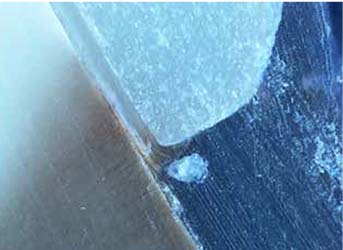
Materials and methods 20 molars were prepared for the placement of lithium disilicate single crowns with vertical margins. Teeth were scanned using a model scanner, in order to create master scans. Then two intraoral scanners (IOS) were used to take impressions of all the 20 prepared teeth: Trios (3 Shape, Copenhagen, Denmark) and Aadva (GC, Tokyo, Japan), so that abutments were scanned with both devices. Then 40 lithium disilicate crowns were fabricated with CAD/CAM technology: each abutment had two crowns made with the two IOS. Then, 20 crowns (10 randomly selected from each IOS group) were luted to the 20 prepared teeth. The crowns were tested for marginal leakage by means of aluminum nitrate solution. Then, teeth were embedded in self-curing transparent resin and cut into 1 mm thick slices by means of a low speed, precision cutting machine (Buehler Isomet) using a diamond blade. The slices of each tooth were observed under optical microscope to evaluate the amount of leakage, if any. Then, the slices were sputter coated with gold and observed under scanning electron microscope (SEM) to evaluate the thickness of the cement at the margins.
Results No statistically significant differences were found, neither regarding the nanoleakage of the crowns made with the two tested IOS nor regarding cement thickness. Measurements of cement thickness were on average within the acceptable limits considered.
Conclusions Both IOS tested showed good performances and, from the results of this in vitro study, can be considered useful for clinical application.


How to Cite

This work is licensed under a Creative Commons Attribution-NonCommercial 4.0 International License.
The Journal of Osseointegration has chosen to apply the Creative Commons Attribution NonCommercial 4.0 International License (CC BY-NC 4.0) to all manuscripts to be published.