Comparison of internal fit of lithium disilicate crowns fabricated with CAD/CAM technology using two different intraoral scanners
All claims expressed in this article are solely those of the authors and do not necessarily represent those of their affiliated organizations, or those of the publisher, the editors and the reviewers. Any product that may be evaluated in this article or claim that may be made by its manufacturer is not guaranteed or endorsed by the publisher.
Authors
Aim The aim of this in vitro study was to compare variations in the internal fit of lithium disilicate single crowns fabricated with computer-aided design and computer-aided manufacturing (CAD/CAM) technology using two digital impression systems.
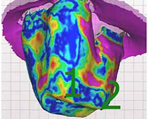
Methods 20 molars were prepared for lithium disilicate single crowns with vertical margins. The teeth were scanned using a model scanner in order to create master scans. Then, two intraoral scanners (IOS) were used to take impressions of the 20 teeth: Trios 3 Basic (3Shape, Copenhagen, Denmark) and Aadva (GC, Tokyo, Japan). The 40 .stl files of the impressions were exported and overlapped with the master scans using the software Aadva GC 2.1.2 Dental DB that, using colors from blue to red, highlights (in red) the areas of discrepancy along the impressions of the abutments. The ratio of red was evaluated to assess if there were any statistically significant differences between the two scanners. The digital impressions were used to fabricate 40 lithium disilicate crowns by means of CAD/CAM technology (for each abutment two crowns were fabricated with both devices). Then, 20 crowns, 10 from each IOS device, were randomly selected and luted to the 20 prepared teeth. Teeth were embedded in self-curing transparent resin and then cut into 1 mm thick slices by means of a low speed, precision cutting machine (Buehler Isomet) using a diamond blade. Slices were then observed under optical microscope (Nikon) to evaluate cement thickness around the abutments.
Results No statistically significant differences were found, regardless of precision discrepancies in the impressions taken with the two tested IOS systems. The marginal fit of complete lithium disilicate crowns made with a complete digital workflow from the impression taken with the two tested devices showed comparable levels of marginal fit.
Conclusions Both intraoral scanners tested showed good performance and, based on the results of this in vitro study, they both can be considered useful for clinical application.
Downloads
Citations
How to Cite

This work is licensed under a Creative Commons Attribution-NonCommercial 4.0 International License.