Surface area coverage of four extra-oral 3D scanning strategies for edentulous arches
All claims expressed in this article are solely those of the authors and do not necessarily represent those of their affiliated organizations, or those of the publisher, the editors and the reviewers. Any product that may be evaluated in this article or claim that may be made by its manufacturer is not guaranteed or endorsed by the publisher.
Authors
Aim The aim of this study was to assess the surface area coverage of four different extra-oral scanning strategies for edentulous arches.
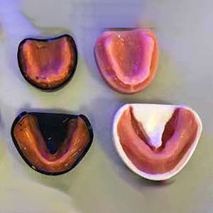
Materials and methods Impressions were taken of six different edentulous models. Gypsum casts were poured from the impressions. The impressions and/or casts were scanned using four different scanning protocols. Three of the protocols used a custom-built 5-axis laboratory scanner; the final protocol used a commercially available 2-axis laboratory scanner (Rexcan DS2). Group imp-5Ax consisted of the scanned impressions, Group cast-5Ax, the scanned the casts, the third group used a “hybrid” method and cast-Ax2 consisted of scans of casts scanned in the laboratory scanner. All scans were repeated five times each to ensure consistency in the data. All scans were uniformly cropped using custom software. Meshlab was used to calculate the surface area coverage obtained from each scanning protocol. Results were compared using ranked ANCOVA and Friedmans test.
Results Overall, there was no significant difference across scanning methods from the ranked ANCOVA test. However, individual nonparametric testing with Bonferroni correction revealed one model differed significantly in surface area (p=0.006) with the hybrid group producing the greatest surface area. The trend showed the hybrid group produced the largest surface area, indicating fewer holes, more frequently than any of the other groups. The commercial 2-axis laboratory scanner was found to produce the smallest surface area most frequently of the four groups, despite being the only group which had undergone hole-filling prior to analysis.
Conclusion Overall, there was no statistical significance between scanning methods, but this does not rule out clinically significant differences in the surface coverage of each scan method. Further studies with a larger sample size would be required to overcome the limitations within this study, but findings indicate a tendency for the hybrid method to produce scans with a larger surface area than all other scan methods investigated.


How to Cite

This work is licensed under a Creative Commons Attribution-NonCommercial 4.0 International License.
The Journal of Osseointegration has chosen to apply the Creative Commons Attribution NonCommercial 4.0 International License (CC BY-NC 4.0) to all manuscripts to be published.