Area accuracy gradient and artificial markers: a three-dimensional analysis of the accuracy of IOS scans on the completely edentulous upper jaw
All claims expressed in this article are solely those of the authors and do not necessarily represent those of their affiliated organizations, or those of the publisher, the editors and the reviewers. Any product that may be evaluated in this article or claim that may be made by its manufacturer is not guaranteed or endorsed by the publisher.
Authors
Aim The present paper aimed to assess the accuracy gradient of scans made using an intraoral scanner (IOS) on a totally edentulous maxilla and the effectiveness of artificial markers.
Materials and methods A reference scan was made by scanning a fully edentulous upper jaw cast (RC) with a dedicated metrological machine. On the RC, an IOS was used to make 10 scans then superimposed to detect their area accuracy gradient. Artificial markers with a diameter of 2 mm were placed in the less accurate areas following two approaches. In the first one, semispherical resin composite markers were used. In the second approach, a dermographic pen was used to draw circular flat markers. Three experimental groups (n = 10) were obtained: “no markers” for the control group without markers, “embossed markers” for resin composite markers, and “flat markers” for ink-drawn ones. The scans were processed into a specialized software, where trueness and precision were measured in millimeters. Descriptive statistics (95% C.I.) were conducted, also, the Games-Howell and Kruskal-Wallis tests (α = .05) were used to investigate differences between groups.
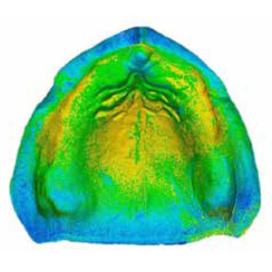
Results Mean values for trueness were: no markers 48.8 (39.2-58.3); embossed markers 39.2 (37.5-40.8); flat markers 60.5 (47.7-73.4), with statistically significant differences between embossed and flat markers (p = .011). Mean values for precision were: no markers 46.7 (29.7-63.7); embossed markers 41.4 (34.7-48); flat markers 99.8 (69.3-130.3), with significant differences between embossed markers and flat ones (p = .008) and between the latter and the control group (p = .005). Minor accuracy was detected at both tuberosities, palate, posterior aspect of the papilla, and flattened areas of the ridges.
Conclusions To improve IOS scans accuracy on the totally edentulous upper jaw, it is suggested to place embossed markers, rather than flat ones, in the areas of minor accuracy.


How to Cite

This work is licensed under a Creative Commons Attribution-NonCommercial 4.0 International License.
The Journal of Osseointegration has chosen to apply the Creative Commons Attribution NonCommercial 4.0 International License (CC BY-NC 4.0) to all manuscripts to be published.