Full-arch intraoral scanning: comparison of two different strategies and their accuracy outcomes
All claims expressed in this article are solely those of the authors and do not necessarily represent those of their affiliated organizations, or those of the publisher, the editors and the reviewers. Any product that may be evaluated in this article or claim that may be made by its manufacturer is not guaranteed or endorsed by the publisher.
Authors
Aim To test if there is a difference in accuracy between full-arch scans performed as two separate halves and stitched together, or as one continuous scan from side to side.
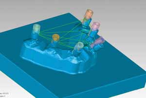
Materials and Methods A reference model with six implants was milled as a single titanium block. Six scan bodies were manufactured and screwed into the implants. A reference 3D model was created using an industrial optical scanner. The experiment was performed using the same intraoral scanning machine (3M True Definition Scanner). The €˜Stitching€™ strategy had the scan started from #27 to #13;after saving this part, the same procedure was performed from #17 to #23 and the software stitched the two halves automatically. The €˜No Stitching€™ strategy had the scan performed as a single procedure. Using engineering software, six copies of the scan body CAD file were substituted to the six scan bodies of the RM and the centre point of each one was determined. Linear measurements were made between the detected points; mean distance and standard deviation were calculated for each of the fifteen measurement sets created.
Results Stitching and No Stitching did not show statistically significant differences (Stitching=0.0396 mm ±0.0409 mm, No Stitching=0.0452 mm ±0,0481 mm, p=.338) but they differed significantly comparing absolute errors (Stitching=0.0442 mm ±0.0358 mm, No Stitching=0.0555 mm±0,036 mm, p=.015).
Conclusions Stitching showed a better precision compared to No Stitching, exhibiting a smaller standard deviation and a higher error density closer to zero.


How to Cite
The Journal of Osseointegration has chosen to apply the Creative Commons Attribution NonCommercial 4.0 International License (CC BY-NC 4.0) to all manuscripts to be published.