Ectopic tooth involving the orbital floor and infraorbital nerve
HTML: 71
All claims expressed in this article are solely those of the authors and do not necessarily represent those of their affiliated organizations, or those of the publisher, the editors and the reviewers. Any product that may be evaluated in this article or claim that may be made by its manufacturer is not guaranteed or endorsed by the publisher.
Authors
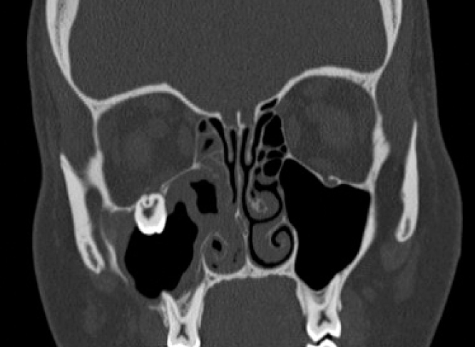
Aim Ectopic eruption of a tooth within the dental arch is often noticed in clinical practice and is well documented in the literature, while ectopic eruption outside the dental arch (as in the maxillary sinus) is rare. Due to its rarity and lack of consensus over management, its incidence needs to be further defined. Surgical approach, with cyst enucleation, has been considered the treatment of choice. In the present study, a case of an ectopic maxillary third molar with chronic purulent maxillary sinusitis is presented. The aim of this report is to describe the extraction of an ectopic third molar carried out through a nasal endoscopic procedure.


How to Cite
The Journal of Osseointegration has chosen to apply the Creative Commons Attribution NonCommercial 4.0 International License (CC BY-NC 4.0) to all manuscripts to be published.